
As a healthcare worker, you are currently struggling with the battle against COVID-19 (Coronavirus) and trying to find the best ways to help treat your patients. However, the novel SARS-CoV-2 virus is simply wreaking havoc on your patients and any useful information that you can use to fight against it will make a difference.
Now, most of the information and literature on COVID-19 are focused on diagnosing and treating the pulmonary system for COVID-19 (Coronavirus) patients. This makes sense, since the SARS (in SARS-CoV-2 virus) stands for “Severe Acute RESPIRATORY Syndrome.” So it becomes easy to be enamored by the pulmonary system. I’ve recently posted about a complete GUIDE on how to use Lung Ultrasound to evaluate for COVID-10 (Coronavirus) which you can read HERE.
However, there is growing evidence that SARS-CoV-2 is an enemy with many faces and skills. While you are just focusing on the respiratory system, it can attack other organs in your distracted state. Because unlike other common respiratory viruses, SARS-CoV-2 can cause significant cytokine storm, increased interleukins, hyperinflammation, and an increased hypercoagulable state. This allows SARS-CoV-2 to cause damage to not just the lungs, but in severe cases, lead to multi-organ failure and death.
So despite maximizing pulmonary management in these patients, we must think about all of the other organs it can affect such as the kidneys, liver, GI, immune system, and especially the heart (STEMI, myocarditis, pericarditis, tamponade, and pulmonary embolism). In a study in JAMA 2020 (Guo et al) they found a greater than 25% chance of myocardial injury among COVID-19 patients!
As we are risking our lives to fight this battle against COVID-19 (Coronavirus) and help defend our patients, we should not feel hopeless, but ought to have as much knowledge as possible against this ruthless and multi-faceted enemy. I’m here and I’m knee-deep in this fight with you, being both an Emergency Medicine and ICU doctor (arguably the two most affected specialties).
In this post, I want to focus on the Cardiac Complications of COVID-19 (Coronavirus) and introduce to you one of the most powerful tools I’ve used over the past decade to help you manage your COVID-19 patients: Point of Care CARDIAC Ultrasound (aka Echocardiography).
After reading this post you will learn about:
- Current Evidence on COVID-19 Cardiac Complications Identified with Ultrasound
- High-Risk Patients for COVID-19 Cardiac Complications
- A Downloadable PDF Algorithm of How to Evaluate for COVID-19 Cardiac Complications
- How to Perform a Focused Cardiac Ultrasound Exam
- How To Quickly Identify Cardiac Pathology for COVID-19
As evidence is increasing on how the SARS-CoV-2 virus is affecting the heart, I urge you to start learning and performing cardiac ultrasound on your sick COVID-19 patients. Unlike lung ultrasound, where you can potentially get the information CXR or CT, you will definitely miss life-threatening cardiac pathology if you just rely on CXR or CT. You will absolutely need to know how to use Cardiac Ultrasound to evaluate for these pathologies. Okay let’s get to it!
- How Does the SARS-CoV-2 virus Affect The Heart?
- Presentation of Cardiac COVID-19 Patients
- Cardiac Ultrasound in COVID-19 Algorithm
- Cases on Cardiac Complications of COVID-19 Patients
- Case 1: Fulminant Myocarditis from COVID-19
- Case 2: Myopericarditis in COVID-19
- Case 3: Cardiac Tamponade from Myopericarditis in COVID-19
- Case 4: Acute Pulmonary Embolism in COVID-19
- Case 5: Acute Unstable Angina and Coronary Artery Occlusion in COVID-19
- Social Media Cardiac Ultrasound and COVID-19 Cases
- How to Start Performing Cardiac Ultrasound in COVID-19
- Cardiac Pathology Image Gallery
How Does the SARS-CoV-2 virus Affect The Heart?
There are a significant amount of COVID-19 patients (>20%) that will have cardiac complications (Guo et al) that leads to increased mortality (Shi et al). Similar cardiovascular complications were also seen in 2003 during the previous SARS epidemic (Yu et al). The exact mechanism is still being studied, but as far as I can tell, the current belief is that SARS-CoV-2 causes cardiac complications through multiple mechanisms including increased cytokine release, hyperinflammation, adrenergic stimulation, and a hypercoagulable state.
This figure from Champman et al in Circulation 2020 sums it up very well:

Champman et al in Circulation 2020
It seems that SARS-CoV-2 is then able to affect the heart and then potentially lead to these cardiac complications:
- Arrhythmias
- Myocardial injury (with troponin elevation)
- STEMI
- Myocarditis with systolic dysfunction
- Pericarditis
- Myopericarditis
- Pericardial effusion +/- Tamponade
- Pulmonary embolism.
Unfortunately all of these cardiac complications are significant and portend significantly increased morbidity and mortality.
Presentation of Cardiac COVID-19 Patients
After going through all of the Cardiac COVID-19 cases I could find (read further down for detailed descriptions of each of these cases), it seems most patients have similar presentations. This is mainly in the form of cardiac chief complaints (significant dyspnea and/or chest pain) with abnormal vital signs: tachycardia and/or hypotension. With hypotension seeming to be the most specific and worrisome presentation. Therefore, it would be especially important that you rule out COVID-19 cardiac-related complications in Hypotensive COVID-19 patients to make sure you are not missing any life-threatening causes.
Cardiac Ultrasound in COVID-19 Algorithm
Based on cardiac pathology that COVID-19 patients may have and their presenting symptoms, I created a Downloadable PDF below to help you start thinking about using cardiac ultrasound to evaluate for cardiac-related complications of COVID-19. I did not include every single echo finding out there, just the main ones that will be most useful to you to quickly access on 2D/B mode. Going over all of the advanced echocardiographic findings is beyond the scope of this post. I will give examples of these cardiac ultrasound pathologies at the end of the post.
Download PDF of Cardiac Ultrasound Evaluation in COVID-19

Like this Post?
Sign Up For POCUS 101 Updates!

Cases on Cardiac Complications of COVID-19 Patients
Below, I will list the case reports of patients from different countries and how they presented from recently published studies as well as social media posts. Many of these cases do not show the cardiac ultrasound images but I will highlight the echocardiographic findings that they put in RED. For a complete list of all POCUS in COVID-19 literature, check out this post HERE.
Case 1: Fulminant Myocarditis from COVID-19
Citation: Coronavirus fulminant myocarditis treated with glucocorticoid and human immunoglobulin. Hu et al Eur Heart J. 2020 Mar 16
Country: China
HPI: A 37-year-old male with no previous medical history who presents with chest pain and dyspnea x 3 days.
Significant Exam Findings: BP 80/50
Labs: Troponin elevated at > 10,000 ng/L, BNP elevated at 21,035 ng/L, + COVID test, all other respiratory virus tests negative.
Radiology: CXR: Enlarged heart with pulmonary congestion. CT chest: R>L Pleural Effusion and ground glass specifications
EKG: Suspected STEMI in (III, AVF)
Cardiac Ultrasound: Enlarged heart with a marked decrease in ventricular systolic function. LV Ejection Fraction significantly decreased at 27%. Trace (2mm) pericardial effusion.


Coronary Angiogram: NEGATIVE (no coronary stenosis)
Final Diagnoses: Fulminant Myocarditis secondary to COVID-19
Treatment: Supportive treatment and: Metholprednisolone (200mg/day, 4 days), Immunoglobulin (20g/day, 4 days), Norepinephrine, Milrinone, piperacillin sulbactam.
Outcome: 1 week later: Normal CXR, EF with normal LV ejection fraction. 3 weeks later: normalized cardiac markers.
Case 2: Myopericarditis in COVID-19
Citation: Cardiac Involvement in a Patient With Coronavirus Disease 2019 (COVID-19). Inciardi et al JAMA Cardiol. 2020 Mar 27.
Country: Italy
HPI: A 53-year-old white female with no previous cardiovascular disease who presents with severe fatigue x 2 days and cough/fever week before.
Significant Exam Findings: BP 90/50, tachycardia (100 b.p.m.), SpO2 98% on room air, and temp of 36.6 degrees celsius.
Labs: Troponin elevated at 240 ng/L, BNP elevated at 5647 ng/L, + COVID-19 test, all other respiratory virus tests negative.
Radiology: CXR: Unremarkable. Cardiac MRI: Diffuse wall thickness, LVEF 35%, pericardial effusion 12mm.
EKG: ST elevation in infero-lateral leads
Cardiac Ultrasound: LV ejection fraction reduced (40%), Grade 1 diastolic dysfunction, Pericardial effusion (11mm) with no tamponade.


Coronary Angiogram: NEGATIVE (no coronary stenosis).
Final Diagnoses: Myopericarditis from COVID-19
Treatment: Dobutamine, diuresis, ASA, hydroxycholoroquine, lopinavir/rigonavir
Outcome: 6 days later: improvement of symptoms, LV ejection fraction improved to 44% and pericardial effusion decreased to 8mm.
Case 3: Cardiac Tamponade from Myopericarditis in COVID-19
Citation: Life-threatening cardiac tamponade complicating myo-pericarditis in COVID-19. Hua et al Eur Heart J. 2020 Mar 30
Country: United Kingdom
HPI: A 47-year-old female with no previous cardiac risk factors who presents with breathlessness, chest pain, dry cough, and subjective fevers.
Significant Exam Findings: BP 80/50, tachycardia (100 bpm)
Labs: Troponin elevated at 253 ng/L, + COVID-19 test, all other respiratory virus tests negative.
Radiology: CXR: Mild pulmonary congestion.
EKG: ST elevation in infero-lateral leads (III, AVF)
Cardiac Ultrasound: Normal LV ejection fraction. 2cm pericardial effusion with cardiac tamponade.


Coronary Angiogram: Angiogram 3 years prior (2017) was NEGATIVE (no coronary stenosis).
Final Diagnoses: Myopericarditis with resulting Cardiac Tamponade secondary to COVID-19
Treatment: Bedside pericardiocentesis under echocardiographic guidance with output of 540ml of serosanguinous fluid.
Outcome: Immediate improvement of hemodynamic parameters.
Case 4: Acute Pulmonary Embolism in COVID-19
Citation: Acute pulmonary embolism and COVID-19 pneumonia: a random association? Danzi et al Eur Heart J. 2020 Mar 30.
Country: Italy
HPI: A 75-year-old female with 10 days of fever and recent-onset dyspnea.
Significant Exam Findings: “Hemodynamicaly stable” (per article)
Labs: Troponin elevated at 3240 ng/L, + COVID-19 test, all other respiratory virus tests negative.
Radiology: CT: Extensive bilateral ground-glass opacifications and pulmonary emboli of bilateral main pulmonary arteries. Lower extremity ultrasound negative for DVT.
EKG: “normal”
Cardiac Ultrasound: Dilated and severely hypokinetic right ventricle with mean PA pressure of 60 mmHg.


Final Diagnoses: Acute Pulmonary Embolism in the setting of COVID-19
Treatment: Low molecular weight heparin, lopinavir/ritonavir, and hydroxychloroquine.
Outcome: N/A. (POCUS 101 editor note: this is unclear if COVID-19 is a cause or just an association of pulmonary embolism in this case).
Case 5: Acute Unstable Angina and Coronary Artery Occlusion in COVID-19
Citation: Exploring personal protection during high-risk PCI in a COVID-19 patient – Impella CP mechanical support during ULMCA bifurcation stenting. Bettari et al JACC: Case Reports March 2020.
Country: Italy
HPI: A 70-year-old male with no previous cardiac risk factors presents with persistent chest pain.
Significant Exam Findings: BP: 100/65, HR 72 bpm, RR 14 per minute, fever >39 degrees celsius.
Labs: Troponin normal at 11 ng/L, + COVID-19 test, all other respiratory virus tests negative.
Radiology: CXR: “interstitial involvement of the lungs”
EKG: left anterior fascicular block and left axis deviation
Cardiac Ultrasound: Left ventricular ejection fraction of 45% with LAD territory showing severe hypokinesis. (*POCUS 101 editor: they are referring to regional wall motion abnormality of the left ventricle, likely the antero-septal region)
Coronary Angiogram: Chronic total occlusion of Right Coronary Artery (RCA) and Distal Left Main Coronary Artery (LMCA) with critical stenosis


Final Diagnoses: Acute Coronary Syndrome (ACS) with critical LMCA stenosis in the setting of COVID-19
Treatment: Stenting of occluded arteries with left ventricular support device (Impella).
Outcome: Regional wall motion abnormalities resolved and patient hemodynamically stable. (POCUS 101 editor note: this is unclear if COVID-19 is a cause or just an association of ACS in this case. But patients who do have increased cardiac risk factors should likely be screened for ACS given the increased hypercoagulable state in COVID-19. See social media cases below with patients with COVID-19 and STEMI as well)
Like this Post?
Sign Up For POCUS 101 Updates!
Social Media Cardiac Ultrasound and COVID-19 Cases
Thanks Dr. Purvi Parwani (cardiologist extraordinaire) on making a twitter page dedicated to Cardiac COVID cases. You can access it HERE. I’ll highlight some cases from social media below:
How to Start Performing Cardiac Ultrasound in COVID-19
I created a series of videos that anyone can use to learn the basics of cardiac ultrasound. You can access the page HERE. I also listed the videos below to obtain the 4 main cardiac views and IVC view you can use to perform a focused cardiac ultrasound:
Parasternal LONG Axis View
Parasternal SHORT Axis View
Apical 4-Chamber View
Subxiphoid View
IVC View
Cardiac Pathology Image Gallery
Examples: RV Dilation, McConnell’s Sign, Decreased LV EF, Tamponade, Dilated IVC
Here are some additional cardiac ultrasounds showing pathology:
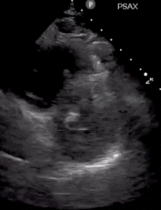

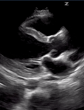

(RV Diastolic Collapse)

(RA Systolic Collapse)

Enjoyed this Guide on Cardiac Ultrasound? Check out these other two posts I made for you on GUIDE to LUNG and MULTIORGAN ULTRASOUND in COVID-19.


[…] POCUS 101 @Vi Dinh @viamdinh Apr 9: Complete Guide to CARDIAC Ultrasound in #COVID19. New Blog POST! What are 5 things you MUST know to … […]